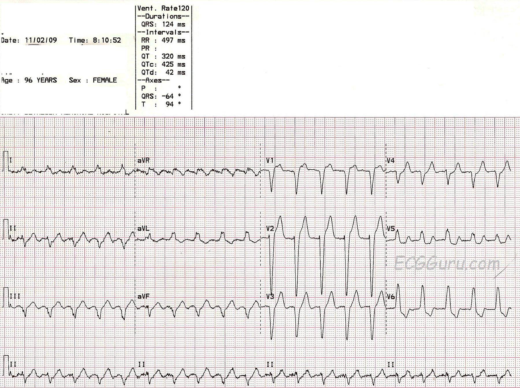

Atrial flutter on ecg full#
The progressive accumulation of accurate activation mapping data of the full spectrum of left atrial reentry will certainly allow the development of clinically useful ECG algorithms. Their morphology can be used to infer the location of the exit from this slow-conducting isthmus and facilitate ablation. As a result, discrete P waves separated by a 12-lead synchronous isoelectric interval may be produced. Although continuous reentrant activation should generate continuous electrical activity on surface ECG, this may not be the case if part of the reentry circuit generates feeble electrical forces, for example, a thin strand of myocardium. Similarly, bystander activation of the right atrium generates a dominantly positive deflection in V1 even during left atrial reentry. Recent studies have shown that nonreentrant arrhythmias originating from the left atrium are characterized by specific ECG features of the P wave, the most consistent being a positive or a dominantly positive deflection in V1. An ideal goal would be to achieve diagnostic precision similar to the ‘sawtooth’ ECG morphology of typical atrial flutter. An improved understanding of ECG manifestations of reentrant left atrial arrhythmias has obvious clinical implications. Atrial flutter is a supraventricular arrhythmia typically seen in patients with underlying. Misdiagnosis of atrial fibrillation occurs more often when atrial activity is prominent on an ECG in more than one lead.The present popularity of catheter ablation for atrial fibrillation has generated interest in the pathophysiology of arrhythmias originating from the left atrium. The hallmark of AF is the irregularly irregular ventricular response. In this lesson, well look at why/how atrial flutter occurs, and then look at a typical ECG readout for a patient in atrial flutter and provide a cardiac interpretation at. When it first occurs, it is usually associated with a fast heart rate.

In conclusion, atrial fibrillation is frequently misdiagnosed as atrial flutter. Atrial flutter (AFL) is a common abnormal heart rhythm that starts in the atrial chambers of the heart.

ECG 3 was correctly identified by 91% of cardiology fellows and cardiologists and by 82% of house officers and internists (P =. ECG 2 was correctly identified by 26% of cardiology fellows and cardiologists and by 37% of house officers and internists (P =. Cardiology fellows and cardiologists correctly identified ECG 1 more often than house officers and internists (95% vs 63% P < or =. Overall, ECG1 was correctly identified as atrial fibrillation by 79% of physicians, ECG 2 was correctly identified as atrial fibrillation by 31%, and ECG 3 was correctly identified as atrial flutter by 90%. ECG 1 showed atrial fibrillation with prominent atrial activity (>0.2 mV) in lead V1 ECG 2 displayed atrial fibrillation with prominent atrial activity (>0.2 mV) in leads III and V1 and ECG 3 displayed atrial flutter. Electrocardiogram Characteristics of Atrial Flutter Regular rhythm with heart rate around divisors of 300 (150 bpm, 100 bpm, 75 bpm). A questionnaire containing three 12-lead ECGs was mailed to 689 physicians, with multiple-choice questions asking whether the rhythm on each ECG was atrial flutter or atrial fibrillation. The purpose of this study was to determine the ability of physicians to differentiate atrial flutter from atrial fibrillation on a surface electrocardiogram (ECG). Atrial flutter (AFl) is a cardiac dysrhythmia characterized by rapid and regular depolarization of the atria that appears as a sawtooth pattern on the electrocardiogram (ECG) and is categorized into type I (typical) and type II (atypical) AFl.


 0 kommentar(er)
